tree in bud radiology assistant
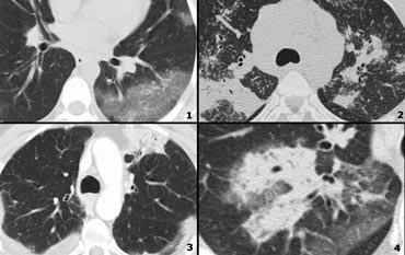
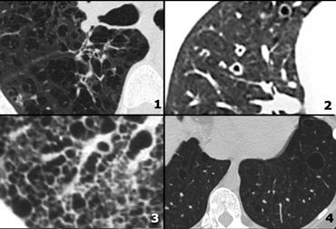
As its name implies this pattern resembles a budding tree in CT scans see Fig. Tree-in-bud describes the appearance of an irregular and often nodular branching structure most easily identified in the lung periphery.
The Radiology Assistant Hrct Basic Interpretation
Pus mucus or inflammatory exudate centrilobular bronchioles.
. The Tree-in-Bud Pattern. Tree-in-bud TIB is a radiologic pattern seen on high-resolution chest CT reflecting bronchiolar mucoid impaction occasionally with additional involvement of adjacent alveoli. 1 From the Department of Radiology University of Vienna Waehringer Guertel 18-20 A-1090 Vienna Austria.
Received November 11 1999. Tree in bud opacification refers to a sign on chest CT where small centrilobular nodules and corresponding small branches simulate the appearance of the end of a branch belonging to a tree that is in bud. Address correspondence to the author e-mail.
3 found that the tree-in-bud pattern was seen in 256 of the CT scans in patients with bronchiectasis. Originally and still often thought to be specific to endobronchial Tb the sign is actually non-specific and is the. Originally reported in cases of endobronchial spread of Mycobacterium tuberculosis this pattern is now.
In centrilobular nodules the recognition of tree-in-bud is of value for narrowing the differential diagnosis. Usually somewhat nodular in appearance the tree-in-bud pattern is generally most pronounced in the lung periphery and associated with abnormalities of the. Cases with TIB opacities in the radiology report in 2010 were identified by searching the Radiology Information System.
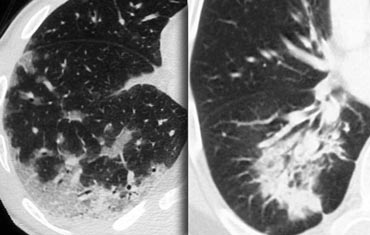
1 refers to a pattern seen on thin-section chest CT in which centrilobular bronchial dilatation and filling by mucus pus or fluid resembles a budding tree Fig. Tree-in-bud appearance represents dilated and fluid-filled ie. Certainly the cause of her symptoms is more likely to be the terminal bronchial plugging with tree in bud.
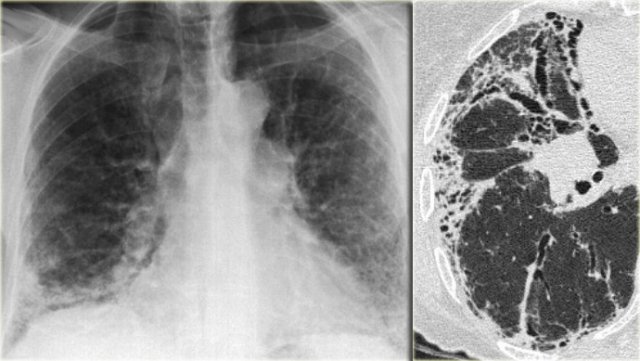
Mollura in Computerized Medical Imaging and Graphics 2012 526 Mixtures tree-in-bud. Multiple causes for tree-in-bud TIB opacities have been reported. The CTPA demonstrates a small peripheral right-sided pulmonary embolus but more significant is the widespread terminal bronchial plugging and bronchial wall thickening.
Bronchial wall thicken-ing is a potentially reversible finding and correlates with. To make it even worse it was bordering on my neighbor s lot and the old man who lived there was even more superstitious than most. Tree-in-bud TIB opacities are a common imaging finding on thoracic CT scan.
Small nodules in a perilymphatic distribution ie. 1-4Reported causes include infections aspiration and a variety of infl ammatory. The other is centrilobular nodules.
Tree-in-bud describes the appearance of an irregular and often nodular branching structure most easily identified in the lung periphery. Fig 7 Tree In Bud Sign Chest Ct Shows Tree In Bud Images Schematic Drawings And Corresponding Picture Refe Radiology Radiology Imaging Medical Radiography. Revision requested December 10.
It is usually pronounced in centrilobular branching structures in the lung periphery associated with diseases of the small airways 36The tree-in-bud sign indicates bronchiolar. However to our knowledge the relative frequencies of the causes have not been evaluated. Cases with TIB in the radiology report in 2010 were identified by searching the Radiology Information System.
J Comput Assist Tomogr 1996. Its microbiologic significance has not been systematically evaluated. The Radiology Assistant Basic Interpretation Idiopathic Pulmonary.
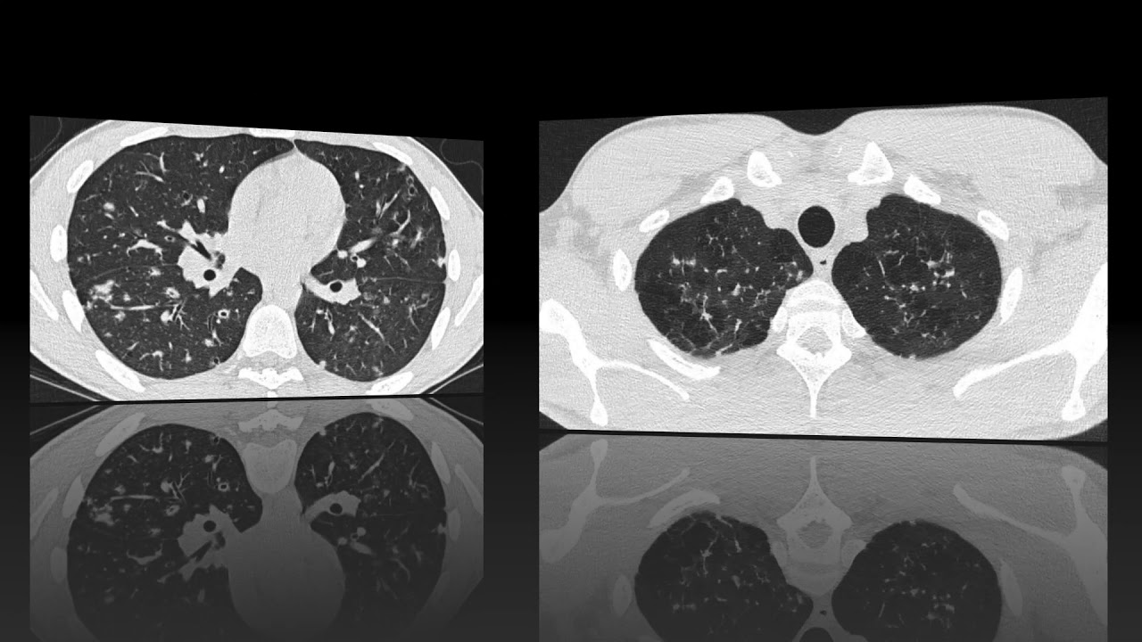
Along subpleural surface and fissures along interlobular septa and the peribronchovascular bundle. Abnormal tree-in-bud bronchioles can be distinguished from normal centrilobular bronchioles by their more irregular appearance lack of tapering or knobbybulbous appearance at the tip of their branches. Tree-in-bud pattern and poorly defined nodules representing bronchiolar filling.
It represents dilated and impacted mucus or pus-filled centrilobular bronchioles. The tree-in-bud pattern was first used as a descriptor by Im et al. To describe the appearance of the endobronchial spread of mycobacterial tuberculosis.
Lingular atelectasis may be a chronic finding. One characteristic feature of bronchiolar disease is a tree-in-bud pattern on computed tomography CT. Normal lobular bronchioles 1 mm in diameter cannot be seen on CT scans which can only show bronchi more than 2 mm in diameter.
Upper and middle zone predominance. It represents dilated and impacted mucus or pus-filled centrilobular bronchioles. Diagnosis Pathophys Radiology Pulmonary CTChest TreeInBud Diagram RadiologyAssistant.
Revision received and accepted May 22 2000. The Tree-in-Bud Sign. We aimed to establish the incidence of the TIB pattern as a proportion of all patients undergoing chest CT.
Tree-in-bud describes the appearance of an irregular and often nodular branching structure most easily identified in the lung periphery. Tree in bud radiology assistant Monday April 4 2022 Edit. The tree-in-bud pattern is commonly seen at thin-section computed tomography CT of the lungs.
31 March 2013. Tree-in-bud pattern simulating diffuse panbronchiolitis but without cylindrical bronchiectasis. Lymphadenopathy in left hilus right hilus and paratracheal 1-2-3 sign.
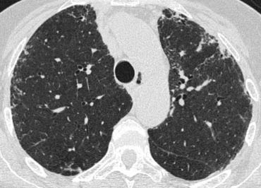
Mar 25 2020 - Poster. Medical records and CT scan examinations. It consists of small centrilobular nodules of soft-tissue attenuation connected to multiple branching linear structures of similar caliber that originate from a single stalk.

Cylindrical Bronchiectasis And Tree In Bud Pattern In Lower Lobes And Download Scientific Diagram

Pin Ot Polzovatelya Emily Shaffer Na Doske Rads Appendix Reference Images Medicinskaya Shkola Radiologiya Medicinskij

Pin On Chest Ct Mri
1
The Radiology Assistant Hrct Common Diagnoses
The Radiology Assistant Hrct Basic Interpretation
The Radiology Assistant Hrct Basic Interpretation

Palla S Sign

Fig 7 Tree In Bud Sign Chest Ct Shows Tree In Bud Images Schematic Drawings And Corresponding Picture Refe Radiology Radiology Imaging Medical Radiography

Pin By Nguyễn Thị On Hrct16 Medical Radiography Radiology Imaging Medical Anatomy

Fig 22 Mosaic Pattern Chest Ct With Schematic Drawing And Illustrative Picture References Radiology Univer Radiology Radiology Imaging Diagnostic Imaging

Bronchiectasis Medical Radiography Radiology Diagnostic Medical Sonography

Learningradiology Lung Abscess Pulmonary Lunges Pulmonary X Ray
Fleischner Sign

Holman Miller Sign Antral Sign
Tree In Bud
The Radiology Assistant Hrct Basic Interpretation
The Radiology Assistant Hrct Common Diagnoses
Hondusa Sign